
Quick Answer: What Causes PUJ Obstruction in Children?
Pelvi Ureteric Junction Obstruction (PUJO) in children is most frequently caused by an intrinsic congenital muscular defect within the junction wall (an aperistaltic segment) that fails to contract and propel urine down the ureter. In older children, it is commonly triggered by external compression from an aberrant crossing lower pole blood vessel. Less frequent causes include structural stenosis, high ureteric insertion, persistent embryonic folds, or acquired scarring from stones and infections. Early evaluation by an experienced pediatric urologist safeguards the child's long-term kidney function.
Receiving an ultrasound report showing a swollen kidney is naturally worrying for parents. Many families first learn about Pelvi Ureteric Junction Obstruction (PUJO)—also referred to internationally as ureteropelvic junction obstruction (UPJO)—during a routine prenatal scan or after an infant develops an unexpected fever or tummy ache. Naturally, parents wonder: What caused this blockage? Did we do something wrong during pregnancy? Will my child's kidney recover?
Under the clinical guidance of renowned pediatric urologist الدكتور سوجيت شودري, this comprehensive medical guide explains the anatomical and developmental causes of Pelvi Ureteric Junction Obstruction in children in clear, simple language, providing the clinical answers you need to make confident decisions for your family.
Understanding the Pelvi-Ureteric Junction
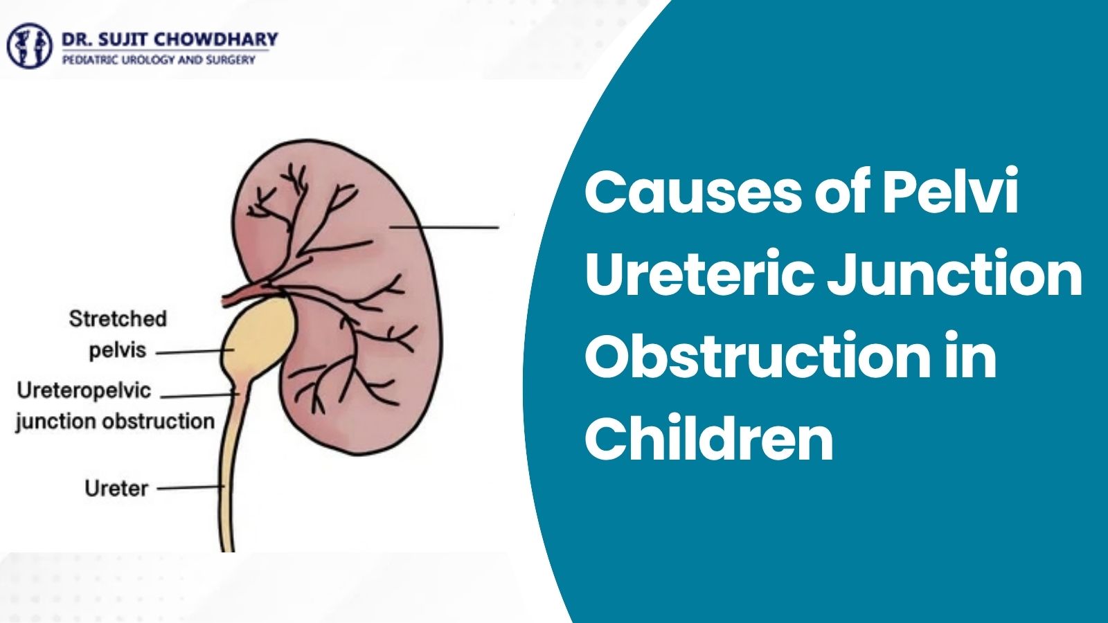
To understand the blockage, it helps to review normal kidney function. The kidneys filter waste and fluid from the blood to produce urine. Inside each kidney, funnel-like collection cups merge into a central chamber called the renal pelvis.
At the lower end of this funnel lies the Pelvi-Ureteric Junction (PUJ). This junction connects the wide renal pelvis to the ureter, the muscular drainage tube that carries urine down to the urinary bladder. Urine does not move by gravity alone; the walls of the junction and ureter contain specialized smooth muscle fibers that create wave-like contractions (peristalsis) to pump urine forward. When this junction is physically narrowed or fails to contract, urine accumulates inside the renal pelvis, creating backpressure that expands the kidney—a condition termed استسقاء الكلى لدى الأطفال.
Concerned About Antenatal Kidney Swelling or PUJO?
Consult with internationally acclaimed pediatric urologist Dr. Sujit Chowdhary for precise diagnostic scans, gentle monitoring, and world-class robotic care in Delhi.
Book a ConsultationPrimary Classifications: Intrinsic vs. Extrinsic Causes
Pediatric urologists classify the causes of Pelvi Ureteric Junction Obstruction in children into two fundamental anatomical categories:
- Intrinsic Causes: Defects originating within the structural wall, muscle tissue, or inner lining of the junction itself.
- Extrinsic Causes: Outside structures, such as crossing blood vessels or abnormal fibrous bands, that press against or kink the junction from the exterior.
The vast majority of childhood cases are congenital (present from birth), while a small minority are acquired later in childhood.
Congenital & Intrinsic Causes (Inside the Ureteral Wall)
Intrinsic developmental factors account for more than 75% to 80% of all PUJ obstruction cases diagnosed during infancy. These occur during early embryonic development between the 4th and 12th weeks of pregnancy:
1. The Aperistaltic (Adynamic) Segment
The most common intrinsic cause of PUJO in infants is an aperistaltic or adynamic segment. In a healthy junction, circular smooth muscles contract in coordinated waves. In affected infants, these muscle fibers are deficient or replaced by non-contractile collagen. Even though the passage appears open, it cannot generate contraction waves. Urine pools above the junction, causing chronic backpressure and pelvic dilation.
2. Congenital Intrinsic Stenosis (Narrowing)
During embryonic development, the ureter solidifies and later recanalizes. If this process is incomplete, a narrow constriction remains at the junction, severely restricting urine flow into the ureter.
3. High Ureteric Insertion
Normally, the ureter exits at the lowest point of the renal pelvis funnel for smooth drainage. In children with high ureteral insertion, the ureter attaches abnormally high up on the wall of the renal pelvis, like a tap placed halfway up a bucket. The pelvis must fill completely under elevated pressure before urine spills over, leading to persistent fluid retention.
4. Mucosal Folds and Embryonic Valves
Temporary mucosal folds and subtle kinks normally smooth out before birth. Occasionally, small valve-like mucosal flaps linger at the junction, acting as one-way barriers that hinder urine outflow.
Clinical Insight for Parents
Congenital intrinsic causes of PUJO are spontaneous developmental occurrences. They are not caused by maternal diet, medications, or everyday activities during pregnancy. With timely pediatric urological oversight, kidney function is safely preserved.
Extrinsic Causes (External Mechanical Compression)
Extrinsic causes involve outside structures that mechanically pinch or distort the pelvi-ureteric junction. They represent the most common cause of PUJO diagnosed in school-aged children and adolescents:
1. Aberrant Crossing Lower Pole Vessels
In 20% to 30% of older children with PUJO, an accessory blood vessel courses across the junction. As the child grows or drinks large amounts of fluids, the expanding pelvis pushes against this taut vessel. The junction is compressed from the outside, causing intermittent obstruction and intense flank pain known as Dietl's crisis.
2. Fibrous Bands and Adhesions
Embryological connective tissues that fail to resorb cleanly form fibrous bands. These tether the upper ureter, pulling it into an acute kink that restricts normal drainage.
3. Associated Congenital Renal Variations
Children born with structural kidney variations have a higher susceptibility to extrinsic PUJ obstruction:
- Horseshoe Kidney: Both kidneys are fused across the midline, forcing the ureters over the lower tissue bridge, predisposing to high insertion and compression.
- Ectopic or Pelvic Kidneys: Kidneys located in the pelvis rather than the upper abdomen, often featuring atypical vessels and acute junction angles.
Intrinsic vs. Extrinsic PUJO: Key Differences
| الميزة | Intrinsic Causes | Extrinsic Causes |
|---|---|---|
| Primary Mechanism | Aperistaltic muscle segment, stenosis, or mucosal folds | Compression by crossing lower pole vessels or fibrous bands |
| Typical Discovery Age | Antenatal ultrasound or early infancy (< 1 year) | Older children (5–15 years) and adolescents |
| Obstruction Pattern | Constant, steady baseline restriction | Intermittent; triggered by heavy hydration or sports |
| Common Signs | Often silent; infant fever (UTI), poor weight gain | Episodic flank pain (Dietl's crisis), vomiting, hematuria |
| Surgical Solution | Resection of narrow segment & Anderson-Hynes Pyeloplasty | Transposition anterior to vessel & Dismembered Pyeloplasty |
Secondary & Acquired Causes in Children
Although rare in pediatric medicine, PUJO can be acquired during childhood through secondary medical conditions:
- التهابات المسالك البولية المتكررة (التهابات المسالك البولية): Severe bacterial infections can produce chronic inflammation that heals with scar tissue, narrowing the junction lumen. Review warning signs in our article on أعراض عدوى المثانة لدى الأطفال.
- Pediatric Kidney Stones (Urolithiasis): A stone lodged at the junction causes mucosal trauma, leading to inflammatory strictures and microscopic or visible red urine, as detailed in our guide on causes of red urine in a child.
- Severe Vesicoureteric Reflux (VUR): Backward urine pressure elongates the ureter, causing tortuosity and secondary kinking at the junction.
Presentation Across Different Age Groups
The clinical presentation of PUJ obstruction varies significantly depending on the child's age:
In Newborns and Infants
Today, over 80% of infant PUJO cases are detected before birth on routine pregnancy ultrasounds. Most newborns have no outward symptoms at birth. When signs do occur, they usually manifest as unexplained fevers, irritability, poor feeding, or a palpable abdominal mass. To understand early indicators, explore our guide on urological problems in newborns.
In Older Children and Teens
Older children typically experience intermittent symptoms driven by crossing vessels or partial narrowings. Hallmarks include recurring bouts of intense flank or tummy pain, nausea and vomiting (often mistaken for appendicitis), pain flaring after heavy fluid intake, and blood in the urine following physical play.
Diagnostic Evaluation: Finding the Underlying Cause
Determining the exact anatomical cause and evaluating whether the kidney is under threat requires specialized pediatric imaging:
- Renal Ultrasound: Measures the renal pelvis diameter, caliceal dilation, and kidney cortical thickness.
- MAG3 / DTPA Diuretic Renogram: A functional nuclear scan using a mild diuretic (furosemide) to measure actual drainage curves and split kidney function percentages.
- MR Urography (MRU) / CT Angiography: Provides high-resolution anatomical mapping of renal blood vessels and collecting systems without ionizing radiation, ideal for detecting crossing vessels.
- Micturating Cystourethrogram (MCU): Excludes associated bladder reflux when clinically indicated.
Gold-Standard Pediatric Urology Care in Delhi
Discover advanced, minimally invasive Pelvi Ureteric Junction Obstruction treatment in Delhi led by Dr. Sujit Chowdhary.
Book an Expert EvaluationTreatment Options: Protecting Renal Function
Management depends on whether the obstruction is impairing kidney drainage or causing functional decline:
1. Active Surveillance
Many infants with mild to moderate hydronephrosis have non-obstructive dilation that improves spontaneously as the urinary tract matures. These children are monitored safely through serial ultrasounds and renograms.
2. Surgical Correction: Dismembered Pyeloplasty
When scans confirm true obstruction, declining renal function, or severe symptoms, surgery is recommended. The gold standard procedure is the Anderson-Hynes Dismembered Pyeloplasty. The surgeon removes the narrowed junction segment, transposes crossing vessels if present, reduces redundant pelvic tissue, and reconstructs a wide, funnel-shaped passage with delicate absorbable sutures.
3. Minimally Invasive & Robotic Pyeloplasty
Under the expertise of master pediatric surgeon الدكتور سوجيت شودري, robotic-assisted pyeloplasty delivers unparalleled precision:
- High-definition 3D visualization of delicate microscopic tissues and blood vessels
- Miniature wristed instruments that replicate human hands with tremor-free accuracy
- Tiny keyhole incisions (5–8 mm) ensuring minimal discomfort and negligible scars
- Short hospital stays (typically 24 to 48 hours) and rapid recovery
- Exceptional success rates exceeding 95% to 98%
Frequently Asked Questions About PUJ Obstruction Causes
What is the most common cause of Pelvi Ureteric Junction Obstruction in babies?
The leading cause in infants is a congenital intrinsic muscular defect known as an aperistaltic segment. The circular muscle fibers at the junction do not develop properly, preventing the peristaltic pumping waves needed to empty urine from the kidney into the ureter.
Is PUJ obstruction genetic or hereditary?
In almost all children, PUJO occurs sporadically as an isolated developmental variation rather than an inherited genetic disorder. Siblings and parents almost always have completely normal kidneys.
Can maternal diet or lifestyle during pregnancy cause PUJO in a fetus?
No. PUJO is not caused by maternal diet, lifestyle, stress, or everyday activities during pregnancy. It is an intrinsic embryonic anatomical development that cannot be prevented by lifestyle changes.
How does an aberrant crossing blood vessel cause PUJO in older children?
An accessory lower pole renal blood vessel can run directly across the junction. As the child grows or drinks large amounts of fluids, the swelling pelvis presses against this taut vessel, causing an intermittent mechanical pinch that produces sudden flank pain.
Can a child outgrow Pelvi Ureteric Junction Obstruction without surgery?
Mild, non-obstructive cases detected on antenatal ultrasound frequently resolve spontaneously as the urinary tract matures. However, true anatomical obstructions with declining kidney function or worsening swelling do not resolve on their own and require pyeloplasty.
What happens if severe PUJO is left untreated in a child?
Untreated severe obstruction causes continuous urine backpressure, leading to progressive thinning of the renal cortex, irreversible loss of kidney function, recurrent kidney infections, and stone formation.
How do doctors decide if surgery is necessary for PUJO?
Pediatric urologists evaluate serial ultrasounds and nuclear diuretic scans (MAG3 or DTPA). Surgery is advised if split renal function drops below 40%, drainage shows prolonged obstruction, or the child suffers recurrent infections and pain.
Where can families receive advanced Pelvi Ureteric Junction Obstruction treatment in Delhi?
Families can consult Dr. Sujit Chowdhary in New Delhi, a renowned pediatric urologist offering world-class robotic and laparoscopic pyeloplasty with international success rates exceeding 95%.
Conclusion: Protecting Your Child's Kidney Health
Learning that your child has Pelvi Ureteric Junction Obstruction can initially feel daunting, but modern pediatric urology offers highly predictable and gentle solutions. Whether caused by an intrinsic muscular defect identified before birth or an extrinsic crossing vessel diagnosed during childhood, early evaluation by a specialist ensures the kidney remains healthy and functional.
With structured monitoring for mild cases and state-of-the-art robotic pyeloplasty for true blockages, children treated for PUJO thrive and lead active, healthy lives. For expert evaluation, scan reviews, and compassionate Pelvi Ureteric Junction Obstruction treatment in Delhi, schedule an appointment with الدكتور سوجيت شودري today.