
Quick Summary for Expectant Parents
Yes, Anorectal Malformation (ARM) can be suspected antenatally during pregnancy. While direct ultrasound visualization of the fetal anus is technically difficult, sonographers can detect indirect antenatal markers such as dilated fetal bowel loops, intraluminal meconium calcifications (enterolithiasis), fetal pelvic cysts, or associated kidney and spine anomalies. When suspected, a specialized fetal MRI and prenatal consultation with a pediatric surgeon ensure a safe, structured post-delivery management plan.
During pregnancy, every routine ultrasound scan is a milestone filled with hope and expectation for parents. Expectant mothers and fathers eagerly await news of their baby’s growth, heartbeat, and overall development. When a routine antenatal ultrasound scan reveals an unexpected finding or suggests a digestive tract anomaly, it can naturally cause immense anxiety and concern.
Among congenital digestive blockages (such as biliary atresia in infants), an Anorectal Malformation (ARM)—historically termed imperforate anus—is one of the conditions parents frequently inquire about during prenatal evaluations. Expectant parents want clear, reassuring answers: Can Anorectal Malformation be diagnosed antenatally before the baby is born? What are the key ultrasound signs, and how should families prepare for delivery?
In this comprehensive medical guide, leading Senior Consultant Pediatric Surgeon & Urologist الدكتور سوجيت شودري provides expert answers regarding the prenatal detection, ultrasound markers, fetal MRI diagnostic techniques, and delivery planning for babies suspected of having an anorectal anomaly. For families seeking specialized fetal counseling and surgical repair, expert Anorectal Malformation Treatment in Delhi provides world-class care from pregnancy through complete childhood recovery.
Has Your Prenatal Scan Raised Suspicion of Fetal Bowel or Pelvic Anomalies?
Early expert counseling gives parents clarity and peace of mind. Schedule a prenatal surgical consultation with Dr. Sujit Chowdhary in Delhi NCR.
Schedule a Prenatal ConsultationWhat is an Anorectal Malformation (ARM)?
To understand whether an anorectal malformation can be detected before birth, it is helpful first to understand what occurs during early fetal development.
An Anorectal Malformation (ARM) is a spectrum of congenital defects that occur during the 4th to 12th weeks of embryonic development. During this early stage, the distal hindgut and urogenital tract split to form separate openings for bowel movement and urination. When this division is incomplete, the baby is born without a normal anal opening or with an abnormally positioned track.
The spectrum of anorectal malformations includes several clinical variations:
- Imperforate Anus: The anus is completely closed or missing, ending in a blind pouch inside the pelvis.
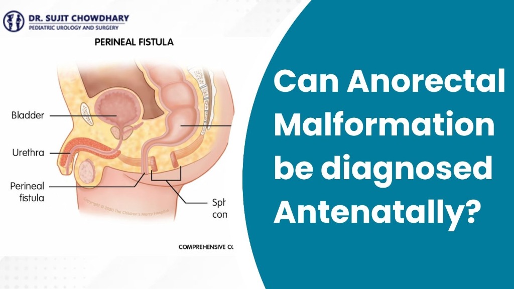
- Perineal Fistula: The bowel opens through a narrow, abnormal tract on the skin close to where the anus should be.
- Rectourethral or Rectovesical Fistula (Boys): The rectum connects directly to the urinary tract (urethra or bladder), allowing meconium to mix with urine.
- Rectovaginal or Rectovestibular Fistula (Girls): The rectum opens into the vaginal vestibule or lower vagina.
- Cloacal Malformation (Girls): A complex malformation where the digestive tract, vagina, and urinary tract merge into a single shared outlet on the perineum.
Medical Fact for Parents
Anorectal malformations occur in approximately 1 in every 4,000 to 5,000 live births worldwide. They affect both boys and girls, ranging from mild low-type anomalies to complex high-type pelvic malformations.
Can Anorectal Malformation Be Diagnosed Antenatally? (Direct Medical Answer)
The direct answer is yes, anorectal malformation can be suspected and diagnosed antenatally during pregnancy, but its detection relies primarily on identifying indirect sonographic signs rather than directly seeing the closed anal opening.
Historically, imperforate anus was considered an exclusively postnatal diagnosis discovered during the routine newborn physical examination immediately after delivery. However, with advances in high-resolution 2D, 3D, and 4D antenatal ultrasound scans and targeted fetal MRI, experienced fetal medicine specialists can now detect subtle prenatal clues during the second and third trimesters.
Scientific studies show that the overall prenatal detection rate for isolated anorectal malformations ranges between 16% and 35%. However, when the condition is associated with complex anomalies (such as cloacal malformations, spinal defects, or renal anomalies), the antenatal detection rate increases to over 60% to 70%.
Why Is Antenatal Diagnosis of ARM So Challenging?
Parents often wonder why a routine ultrasound might not immediately detect an imperforate anus. Sonographers face several anatomical and technical factors during pregnancy:
- Deep Pelvic Location: The fetal anal sphincter and rectum are located deep within the narrow fetal bony pelvis, shaded by acoustic shadows from the fetal pelvic bones.
- Absence of Bowel Gas: Before birth, a fetus does not swallow air. The fetal intestine is filled only with fluid and meconium, so bowel loops do not expand with air as they do after birth.
- Collapse of Distal Rectum: In healthy fetuses, the rectum remains mostly collapsed or empty until late in the third trimester.
- Small Size of Fistulas: Abnormal communication tracts (perineal or rectourinary fistulas) are often microscopic or just 1 to 2 millimeters wide, making direct visualization extremely difficult on routine ultrasound.
Key Antenatal Ultrasound Markers for Anorectal Malformations
When an expert sonographer performs a detailed anomaly scan (level-II ultrasound) or a targeted third-trimester scan, they look for specific indirect markers that hint at distal bowel obstruction or pelvic tract anomalies:
1. Fetal Bowel Dilation (Dilated Distal Colon)
As pregnancy progresses into the third trimester (after 26 to 28 weeks), the fetus ingests amniotic fluid. If the anus is closed, meconium accumulates in the lower colon, causing progressive dilation of distal bowel loops (typically measuring greater than 7 to 10 mm in diameter).
2. Intraluminal Meconium Calcifications (Enterolithiasis)
This is one of the most specific antenatal signs of an anorectal malformation in male fetuses. When a rectourinary fistula connects the rectum to the fetal bladder or urethra, urine mixes with meconium. The chemical reaction causes calcium salts in urine to precipitate, forming bright, echogenic calcified spots (enteroliths) inside dilated fetal bowel loops on ultrasound.
3. Fetal Pelvic Cysts / Hydrocolpos (In Female Fetuses)
In female fetuses with a cloacal malformation, urine and vaginal secretions cannot drain properly and back up into the vagina and uterus. This appears on ultrasound as a large, fluid-filled cystic structure in the fetal pelvis (hydrocolpos), often compressing the bladder and ureters.
4. Absent Perineal Groove or "Target Sign"
In specialized centers, sonographers perform dedicated perineal scanning. A normal fetal anus appears as a central echogenic dot surrounded by a hypoechoic ring of sphincter muscle (the "target sign"). Non-visualization of this sphincter complex or an abnormal perineal contour strongly suggests an anorectal anomaly.
5. Amniotic Fluid Variations (Polyhydramnios)
While polyhydramnios (excess amniotic fluid) is more common in upper GI obstructions (like esophageal atresia), severe low bowel obstruction or associated malformations can occasionally cause mild polyhydramnios due to impaired fetal fluid absorption.
Detailed Prenatal Evaluation & Counseling
If your sonographer has noted fetal bowel dilation or a pelvic cyst, early consultation with a pediatric surgeon provides clear answers.
Explore Anorectal Malformation CareComparison of Diagnostic Techniques for Antenatal ARM Detection
Different diagnostic modalities play distinct roles in evaluating suspected fetal anorectal malformations during pregnancy:
| Diagnostic Tool | Primary Capabilities in ARM Detection | Limitations |
|---|---|---|
| 2D Anomaly Ultrasound | Screens for fetal bowel dilation, renal anomalies, spinal defects, and amniotic fluid volume. | Limited direct visualization of anal sphincter; missed isolated low-type fistulas. |
| 3D / 4D Perineal Ultrasound | Evaluates the anal sphincter target sign, perineal skin fold contour, and pelvic floor muscles. | Requires specialized expertise and favorable fetal positioning (breech or deep pelvis limits view). |
| Fetal MRI (T2-Weighted) | Provides high-contrast images of fetal pelvic organs, meconium high signal, pelvic muscle complex, and spinal cord. | Typically performed after 22–24 weeks; higher cost and requires specialized pediatric radiologist. |
| Postnatal Physical Exam | Immediate 100% confirmation of perineal anatomy, fistula opening, and anal patency within minutes of birth. | Diagnostic only after delivery; does not allow prenatal surgical planning. |
The Role of Fetal MRI in Prenatal Diagnosis
When routine ultrasound raises suspicion of an anorectal malformation or pelvic cyst, Fetal Magnetic Resonance Imaging (MRI) is the gold-standard confirmatory imaging technique.
Fetal MRI uses strong magnetic fields and radio waves without any radiation, making it completely safe for both mother and baby during the second and third trimesters. Because fetal meconium naturally displays a bright signal on T1-weighted MRI sequences, radiologists can track the exact pathway of the distal colon down to the pelvic floor.
Fetal MRI offers several distinct advantages:
- Pinpoints the level of the rectal pouch (high, intermediate, or low ARM).
- Delineates complex cloacal malformations and hydrocolpos in female fetuses.
- Evaluates the pelvic levator ani muscle complex essential for future bowel continent repair.
- Screens for associated spinal cord abnormalities, such as tethered cord or sacral agenesis.
Associated Congenital Anomalies: The VACTERL Association
When an anorectal malformation is suspected antenatally, pediatric specialists immediately perform a thorough check for associated structural issues. Early screening for urological problems in newborns is essential, as over 50% of ARM cases occur alongside other organ anomalies under the VACTERL association:
- V - Vertebral Anomalies: Sacral hemivertebrae or spinal cord anomalies.
- A - Anorectal Malformation: Imperforate anus or fistula.
- C - Cardiac Defects: Ventricular septal defect (VSD) or tetralogy of Fallot.
- TE - Tracheoesophageal Fistula: Abnormal connection between food pipe and windpipe.
- R - Renal / Kidney Anomalies: Single kidney, kidney duplication, or Hydronephrosis in Children.
- L - Limb Differences: Radial ray defects or thumb anomalies.
What Should Expectant Parents Do If an Antenatal ARM is Suspected?
Receiving news that your unborn baby may have an anorectal anomaly can feel overwhelming. However, understanding that modern pediatric surgery offers excellent outcomes helps parents approach delivery with confidence.
Recommended steps for expectant parents include:
- Seek Prenatal Pediatric Surgical Counseling: Schedule a consultation with an experienced pediatric surgeon like الدكتور سوجيت شودري to review ultrasound scans, discuss post-birth expectations, and understand surgical options.
- Plan Delivery at a Tertiary Care Hospital: Ensure your delivery takes place at a medical center equipped with an advanced Neonatal Intensive Care Unit (NICU) and an on-site pediatric surgical team.
- Fetal Echocardiogram & Targeted Ultrasound: Complete a fetal echo and detailed kidney ultrasound to check for associated VACTERL features.
- Reassurance Regarding In-Utero Safety: Anorectal malformations do not harm the fetus inside the womb because waste products are cleared through the placenta into the mother's circulation. Your baby is completely safe until birth.
Postnatal Diagnosis and Immediate Treatment Plan
Once your baby is delivered, the medical team carries out a gentle, structured newborn evaluation:
1. Immediate Clinical Newborn Examination
Within minutes of delivery, the pediatric surgeon examines the baby's perineum to verify the anal opening, check sphincter tone, and check for tiny fistula openings in the perineum, urinary tract, or vagina.
2. Postnatal Imaging (Cross-Table X-Ray & Ultrasound)
After 18 to 24 hours of birth (allowing swallowing of air to outline the bowel), a specialized cross-table X-ray (invertogram) measures the exact distance between the distal rectal pouch and the skin surface. Abdominal and spinal ultrasounds screen the kidneys and spinal cord.
3. Definitive Surgical Reconstruction
Surgical treatment depends on the specific type of anorectal malformation:
- Low-Type ARM / Perineal Fistula: A single, gentle procedure called a primary minimal Posterior Sagittal Anorectoplasty (PSARP) or anoplasty opens the fistula and moves the anus into the center of the muscle sphincter complex.
- High-Type ARM / Rectourinary Fistula / Cloaca: A staged approach is performed. A temporary, life-saving colostomy is created on day 1 or 2 of life to allow normal feeding. Once the baby grows (around 3 to 6 months of age), definitive PSARP or laparoscopic reconstruction is performed by الدكتور سوجيت شودري.
| ARM Type | Postnatal Surgical Approach | Expected Bowel Control Outcome |
|---|---|---|
| Low Perineal Fistula | Single-stage Anoplasty / PSARP in newborn period | Excellent (over 95% full continence) |
| Intermediate / High ARM | Staged approach: Newborn Colostomy → Definitive PSARP at 3–6 months | Good to Excellent with bowel management program |
| Cloacal Malformation | Multidisciplinary reconstruction of bowel, vagina, and urethra | Good with specialized long-term urological and bowel follow-up |
الأسئلة الشائعة (FAQs)
Q1. Can an ultrasound scan detect imperforate anus before birth?
Yes, an antenatal ultrasound scan can raise suspicion of an imperforate anus or anorectal malformation (ARM), primarily by identifying indirect signs such as dilated fetal bowel loops, intraluminal meconium calcification (enterolithiasis), fetal pelvic cysts, or associated kidney and spine anomalies. Direct visualization of the anal dimple is challenging, but indirect markers prompt early expert evaluation.
Q2. At what week of pregnancy can anorectal malformation be suspected?
Anorectal malformation is usually suspected during the mid-trimester anomaly scan (level-II ultrasound) between weeks 18 and 22 of pregnancy. As the fetus grows and produces more meconium in the third trimester (28-34 weeks), bowel dilation or calcified meconium becomes more evident on ultrasound.
Q3. Why is ARM often missed during routine antenatal anomaly scans?
Routine antenatal ultrasound focuses on major organ structures like the brain, heart, spine, and kidneys. The anal sphincter and anal dimple are small soft-tissue structures deep within the fetal pelvis, making them difficult to image directly. Additionally, because fetal intestine does not contain gas before birth, distal bowel dilation may remain subtle.
Q4. Is fetal MRI safe and accurate for diagnosing anorectal malformations?
Yes. Fetal Magnetic Resonance Imaging (MRI) uses magnetic fields without radiation and is entirely safe during the second and third trimesters of pregnancy. It provides high-resolution soft-tissue contrast of the fetal pelvis, helping pediatric surgeons evaluate bowel position, pelvic muscle complex, and rule out spinal cord anomalies.
Q5. Does an antenatal ARM diagnosis mean a high-risk delivery is required?
Antenatal suspicion of ARM does not necessarily require a Caesarean section, but delivery should be planned at a tertiary medical center equipped with a Neonatal Intensive Care Unit (NICU) and an experienced pediatric surgical team for immediate post-birth evaluation.
Q6. Can anorectal malformation be treated before the baby is born?
No fetal or in-utero surgery is performed for anorectal malformations. Treatment is planned immediately after birth. Prenatal identification allows pediatric surgeons to prepare a tailored post-delivery surgical strategy.
Q7. What is the success rate of surgery for children with anorectal malformations?
With modern surgical techniques such as Posterior Sagittal Anorectoplasty (PSARP) and minimally invasive keyhole procedures, over 90% of children with low or intermediate ARM achieve excellent bowel control and live healthy, active lives.
Q8. Where can expectant parents consult for expert Anorectal Malformation Treatment in Delhi?
Expectant parents can consult الدكتور سوجيت شودري, a world-renowned Senior Consultant Pediatric Surgeon & Urologist in Delhi NCR with over 30 years of specialized experience in reconstructive pediatric surgery and prenatal anomaly counseling for Anorectal Malformation Treatment in Delhi.
الخاتمة
In summary, while direct visualization of an imperforate anus remains technically challenging during routine prenatal ultrasound, Anorectal Malformation can definitely be diagnosed or suspected antenatally through indirect sonographic markers such as dilated fetal bowel, intraluminal meconium calcifications, pelvic cysts, and associated anomalies.
Early identification during pregnancy allows expectant parents to meet with a specialized pediatric surgeon, understand what to expect at delivery, and ensure their newborn receives immediate, high-quality care upon birth. With expert surgical reconstruction, children born with anorectal malformations look forward to normal digestion, excellent bowel function, and a bright, healthy future.
Consult Senior Pediatric Surgeon Dr. Sujit Chowdhary
If your prenatal ultrasound scan indicates fetal bowel or pelvic anomalies, seek expert pediatric surgical counseling today.
Book Your Prenatal Consultation